
Calcium Deficiency (Hypocalcaemia): Symptoms, Causes, Diagnosis, Treatment and Prevention -The Complete Doctor’s Guide.
The mineral your bones hold in silence, and what happens when your blood runs low
✍️ Written and reviewed by: Dr. Qazi Taqweemulhaq, FCPS Medicine. Professor of Medicine, Women Medical and Dental College, Abbottabad, Pakistan. Consultant Physician, 32 Years Clinical Experience
📅 Last Updated: May 2026 | References: NIH ODS, NCBI StatPearls, Cleveland Clinic, Merck Manual, Medscape, WHO, UPMC 2025
⚡ Quick Answer: Calcium deficiency (hypocalcaemia: adjusted serum calcium < 2.20 mmol/L) causes muscle cramps, tingling around the mouth and fingertips, Chvostek’s sign, Trousseau’s sign, tetany, and in severe cases, seizures and cardiac arrest. The most common causes are hypoparathyroidism (especially post-thyroid surgery) and Vitamin D deficiency. Always correct the albumin-adjusted calcium, not the raw value. CRITICAL: refractory hypocalcaemia that does not correct with calcium supplementation is almost always caused by concurrent magnesium deficiency, correct magnesium first
|
✅ KEY TAKEAWAYS — Calcium Deficiency |
|
|
|
|
|
|
|
|
• The most common causes of hypocalcaemia are hypoparathyroidism (especially post-thyroid surgery) and Vitamin D deficiency |
|
|
• Always correct calcium with the ‘albumin adjustment’ formula: low albumin gives a falsely low total calcium reading |
|
|
• Severe acute hypocalcaemia (tetany, seizures, QT prolongation) is a medical emergency. IV calcium gluconate required immediately |
🏥 From My Clinic: A 55-year-old woman was referred to me six weeks after a total thyroidectomy for thyroid cancer. She had been discharged from the surgical ward with no calcium supplementation.
She came to me with
progressively worsening hand cramps, tingling around her mouth, and two episodes in which her hands had locked into a characteristic claw-like position, carpopedal spasm.
Her adjusted serum calcium was 1.68 mmol/L, severely low. Her PTH was undetectable.
She had permanent hypoparathyroidism from inadvertent removal of all four parathyroid glands during surgery.
I started IV calcium gluconate followed by oral calcium carbonate and calcitriol.
Within 24 hours she was symptom-free.
She has been on lifelong calcium and active Vitamin D ever since.
I see this pattern regularly in post-menopausal women referred to me with their first fracture.
- A wrist fractured by a minor fall.
- A vertebra collapsed during routine lifting.
And when we look back at years of clinic letters
- Vitamin D levels never checked,
- calcium intake never assessed,
- bone density never scanned
the story tells itself.
But calcium deficiency is not only a slow, silent bone disease. Acute hypocalcaemia (tetany) is one of the most dramatic presentations in internal medicine.
In tetany, there are severe muscular spasms of the hands and feet. Patients can also have painful choking or laryngospasm, and seizures. This dramatic experience is one of medicine’s most genuine emergencies.
Calcium is the mineral that
- regulates every muscle contraction,
- every nerve impulse,
- and every heartbeat.
When serum Calcium level falls precipitously, the consequences can be life-threatening within hours.
This article covers both faces of calcium deficiency:
- the silent, insidious dietary insufficiency that leads to osteoporosis,
- and the acute clinical syndrome of hypocalcaemia that demands immediate treatment.
Understanding both is essential.
What Is Calcium and What Does It Do?
Calcium is the most abundant mineral in the human body, an adult contains approximately 1,000 grams of calcium:
- 99% stored in bone and teeth providing structural strength as hydroxyapatite crystals
- < 1% in blood, muscle, and other tissues but this tiny fraction controls critical physiological functions
The body regulates serum calcium within an extraordinarily narrow range (2.20–2.60 mmol/L) through a sophisticated hormonal system involving:
- Parathyroid hormone (PTH): the primary calcium-raising hormone released when calcium falls, stimulating bone resorption, renal calcium reabsorption, and Vitamin D activation
- Calcitriol (active Vitamin D): enhances intestinal calcium absorption and bone calcium mobilisation
- Calcitonin: released when calcium rises inhibits bone resorption (less clinically significant than PTH)
The Eight Critical Functions of Calcium
- Bone and teeth structure: calcium phosphate as hydroxyapatite provides the rigid mineral matrix of bone. Bones are not inert, they are constantly being remodelled, releasing and depositing calcium throughout life.
- Muscle contraction: calcium is the trigger for every muscle contraction; cardiac, skeletal, and smooth muscle. Without adequate calcium, muscles become hyperexcitable (tetany) or fail to contract properly.
- Nerve impulse transmission: calcium regulates the threshold for nerve firing and is required for neurotransmitter release at the synapse.
Low calcium makes nerves fire spontaneously producing the paraesthesiae, cramps, and tetany of hypocalcaemia.
- Cardiac rhythm: calcium is essential for the cardiac action potential.
- Hypocalcaemia prolongs the QT interval and can cause ventricular arrhythmias and cardiac arrest.
- Blood coagulation: calcium (Factor IV) is required at multiple steps in both the intrinsic and extrinsic clotting cascades.
Severe hypocalcaemia theoretically impairs coagulation, though clinically significant bleeding is rare.
- Cell signalling: intracellular calcium acts as a second messenger for dozens of hormonal and neural signals including those controlling cell division, secretion, and immune activation.
- Enzyme activation: calcium is a cofactor for numerous enzymes throughout the body
- Hormone secretion: calcium regulates the secretion of insulin, growth hormone, and other key hormones
Calcium Blood Test: The Albumin Adjustment – The Most Important Clinical Rule
Here is a clinical rule that every doctor and medical student must know, and that is violated in clinical practice more often than it should be:
Never interpret a total serum calcium result without adjusting for serum albumin.
Approximately 40–45% of serum calcium is bound to albumin.
Total calcium measures both bound and free calcium. In patients with low albumin (hypoalbuminaemia), which is extremely common in hospitalised, malnourished, or chronically ill patients the total calcium appears low even though the biologically active ionised (free) calcium is normal.
The adjustment formula: Adjusted calcium = Measured calcium + 0.02 × (40 − albumin in g/L)
Example: Serum calcium = 1.95 mmol/L; Serum albumin = 20 g/L → Adjusted calcium = 1.95 + 0.02 × (40−20) = 1.95 + 0.40 = 2.35 mmol/L…… NORMAL.
💡 Clinical Insight: Treating apparent hypocalcaemia in a hypoalbuminaemic patient without checking the albumin-adjusted value is one of the most common errors in hospital medicine.
Always calculate the adjusted calcium or request an ionised calcium measurement before starting treatment.
|
Adjusted Serum Calcium |
Status |
Clinical Implication |
|
2.20–2.60 mmol/L (8.8–10.4 mg/dL) |
Normal |
Adequate calcium. No treatment needed for calcium alone |
|
2.00–2.19 mmol/L (8.0–8.8 mg/dL) |
Mild deficiency |
Often asymptomatic. Identify and treat the underlying cause. Oral calcium and Vitamin D supplementation |
|
1.75–1.99 mmol/L (7.0–8.0 mg/dL) |
Moderate deficiency |
Symptomatic in most patients, tetany, cramps, paraesthesiae. Oral supplementation; IV if symptomatic or oral not tolerated |
|
< 1.75 mmol/L (< 7.0 mg/dL) |
Severe deficiency |
Medical emergency. Risk of laryngospasm, seizures, cardiac arrhythmia, cardiac arrest. IV calcium gluconate urgently required |
Table 1. Adjusted serum calcium level interpretation. Sources: NCBI StatPearls; Cleveland Clinic; Merck Manual.
How Much Calcium Do You Need Per Day?
Calcium requirements are highest during adolescence (bone mass accumulation), pregnancy, lactation, and older age (bone loss prevention). The Upper Limit applies to total calcium from all sources, food and supplements combined.
|
Life Stage / Group |
RDA (mg/day) |
Upper Limit (mg/day) |
|
Infants 0–6 months |
200 mg (AI) |
1,000 mg |
|
Infants 7–12 months |
260 mg (AI) |
1,500 mg |
|
Children 1–3 years |
700 mg |
2,500 mg |
|
Children 4–8 years |
1,000 mg |
2,500 mg |
|
Children 9–18 years |
1,300 mg |
3,000 mg |
|
Adults 19–50 years |
1,000 mg |
2,500 mg |
|
Women 51–70 years |
1,200 mg |
2,000 mg |
|
Men 51–70 years |
1,000 mg |
2,000 mg |
|
Adults > 70 years |
1,200 mg |
2,000 mg |
|
Pregnant / Lactating (19–50 yrs) |
1,000 mg |
2,500 mg |
Table 2. Calcium RDA and Tolerable Upper Intake Levels. Source: NIH ODS.
💡 Clinical Insight: Adults absorb only 30–40% of dietary calcium under normal conditions. Absorption efficiency declines with age and is impaired by Vitamin D deficiency, high phytate intake, and low stomach acid. This is why the RDA is set higher than the body’s actual daily requirement.
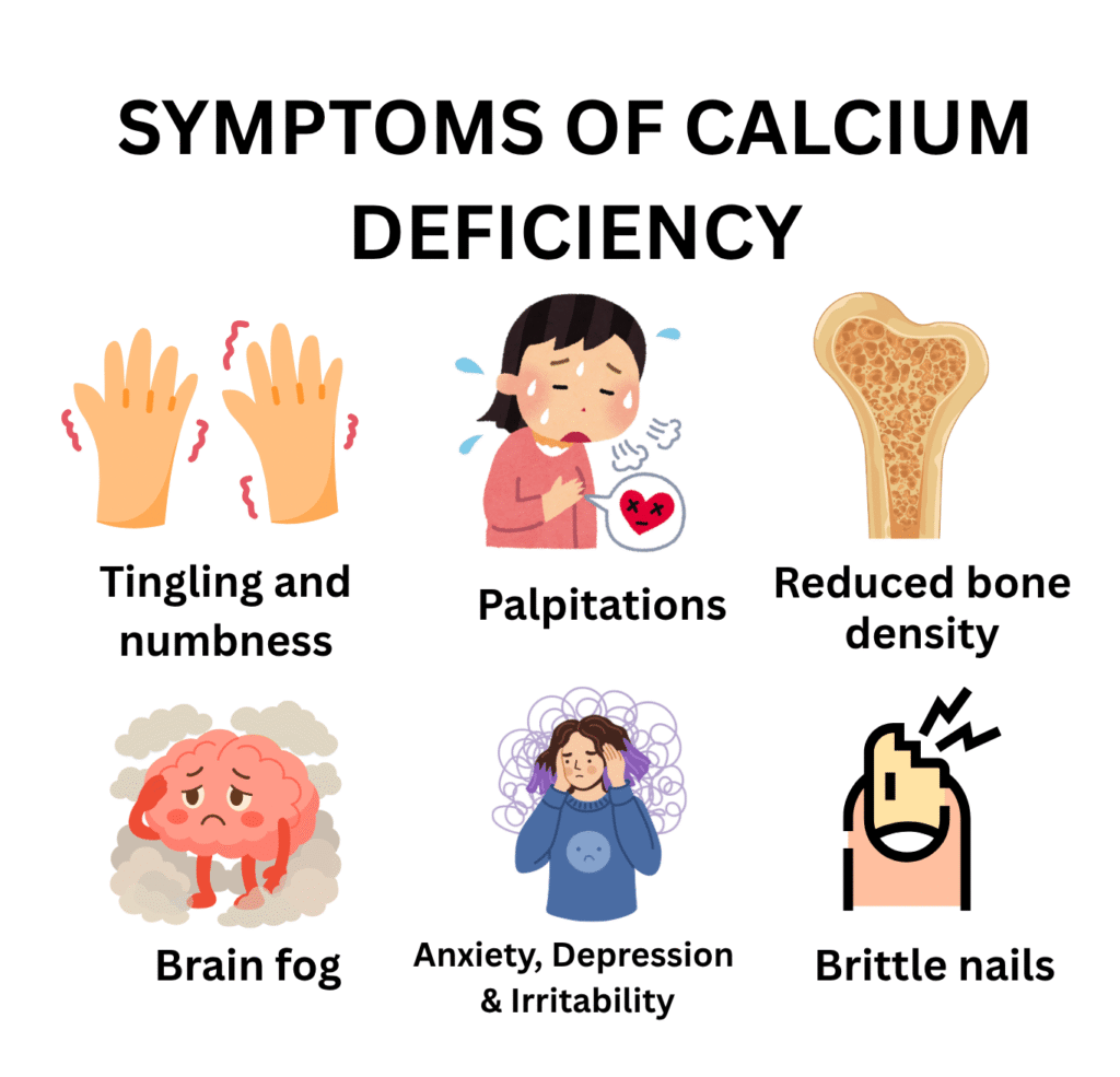
Calcium Deficiency Symptoms: From Tingling to Tetany
The symptoms of calcium deficiency depend critically on how fast and how much calcium falls, not just the absolute level.
A calcium of 1.90 mmol/L that developed slowly over months may be asymptomatic; the same level reached acutely over hours after thyroid surgery may cause life-threatening tetany.
|
System |
Mild–Moderate Deficiency |
Severe Hypocalcaemia |
|
Neuromuscular |
Tingling and numbness (perioral, fingertips, feet), muscle cramps |
Tetany, carpopedal spasm, laryngospasm, generalised seizures |
|
Cardiovascular |
Palpitations |
Prolonged QT interval, ventricular arrhythmia, hypotension, cardiac arrest |
|
Bone (long-term dietary deficiency) |
Osteopenia, reduced bone density without fracture |
Osteoporosis: increased fracture risk; stress fractures; vertebral collapse |
|
Neuropsychiatric |
Brain fog, anxiety, depression, irritability |
Confusion, dementia-like picture, extrapyramidal symptoms (basal ganglia calcification) |
|
Skin, Hair & Nails |
Dry skin, brittle nails |
Coarse dry hair, alopecia, eczematous dermatitis |
|
Eyes |
Mild visual changes |
Subcapsular cataracts: classic complication of chronic hypocalcaemia |
|
Teeth |
Dental decay, poor enamel formation |
Hypoplastic teeth, delayed dentition, dental caries |
|
Children (Rickets) |
Irritability, delayed walking |
Rachitic rosary, bowed legs, growth failure: same picture as Vitamin D deficiency rickets |
Table 3. Calcium deficiency symptoms by severity. Sources: NIH ODS; NCBI StatPearls; Cleveland Clinic; Merck Manual.
The Most Important Symptoms in Clinical Detail
1. Paraesthesiae – The Earliest Symptom
The earliest symptom of hypocalcaemia is characteristically perioral, tingling around the mouth, lips, and tongue, followed by tingling in the fingertips and feet. Patients often describe it as:
- ‘My lips feel numb’
- ‘My hands feel like they’re asleep’
- A burning or prickling sensation around the mouth and face
This pattern of
perioral paraesthesiae followed by extremity tingling is highly characteristic of hypocalcaemia; It should prompt immediate calcium measurement.
2. Muscle Cramps and Tetany
As calcium falls further, neuromuscular hyperexcitability increases:
- Muscle cramps: painful, involuntary muscle contractions, often in the calves and hands
- Tetany: spontaneous tonic-clonic muscle spasms, typically beginning in the hands and feet
- Carpopedal spasm: characteristic flexion of the wrist and extension of the fingers (‘main d’accoucheur’ obstetrician’s hand ) pathognomonic for hypocalcaemia
- Laryngospasm: the most dangerous muscular complication, sudden spasm of the vocal cords causing stridor, respiratory distress, and potentially asphyxia
3. Seizures
Severe or acute hypocalcaemia can cause generalised tonic-clonic seizures clinically indistinguishable from epilepsy.
The key differentiating feature: these seizures resolve completely when calcium is corrected.
Any patient with new-onset seizures should have serum calcium measured as part of the routine workup.
⚠️ Warning: Hypocalcaemia-induced seizures
do not respond to anticonvulsant medications, they respond to IV calcium.
Giving anticonvulsants without correcting the calcium is both ineffective and dangerous.
4. Cardiac Complications
Calcium is essential for the cardiac action potential. Hypocalcaemia causes:
- Prolonged QT interval the most consistent ECG finding; visible on 12-lead ECG
- Ventricular arrhythmia: torsade de pointes and other dangerous rhythms
- Cardiac arrest : in severe, acute hypocalcaemia
- Hypotension and reduced cardiac contractility
💡 Clinical Insight: Always request an ECG alongside the serum calcium in a patient with symptomatic hypocalcaemia.
A prolonged QT interval confirms the cardiac impact and increases the urgency of treatment.
5. Chvostek’s Sign and Trousseau’s Sign – The Bedside Examination
Two bedside signs confirm neuromuscular hyperexcitability from hypocalcaemia:
Chvostek’s sign: tap the facial nerve 2 cm anterior to the ear (just below the zygomatic arch).
A positive sign, twitching of the ipsilateral facial muscles indicates hypocalcaemia.
Note: positive in approximately 10% of normocalcaemic individuals; a negative result does not exclude hypocalcaemia.
Trousseau’s sign: inflate a blood pressure cuff above systolic pressure on the upper arm for 3 minutes.
A positive sign, carpopedal spasm (main d’accoucheur: wrist flexion and finger extension) confirms latent tetany.
More specific than Chvostek’s positive in > 94% of hypocalcaemic patients.
💡 Clinical Insight: Trousseau’s sign is more reliable than Chvostek’s sign. When uncertain, perform Trousseau’s before requesting an urgent ionised calcium. A positive Trousseau’s in a post-thyroidectomy patient is an emergency treat immediately.
6. Osteoporosis – The Silent Long-Term Consequence
When dietary calcium is persistently inadequate over years even without serum calcium falling below normal the skeleton is gradually demineralised as the body reabsorbs bone calcium to maintain serum levels. The result:
- Osteopenia: reduced bone mineral density (T-score between −1.0 and −2.5 on DEXA scan)
- Osteoporosis: severely reduced bone mineral density (T-score below −2.5) with markedly increased fracture risk
- Fragility fractures: fractures occurring from minor trauma: wrist, hip, vertebral, and rib fractures the most devastating clinical consequence of long-term calcium deficiency
Approximately 200 million women worldwide have osteoporosis. Hip fractures, the most serious fragility fracture, have a 20–30% mortality rate within one year in elderly patients.
What Causes Calcium Deficiency? Every Cause Explained
|
Cause |
Specific Cause |
Key Mechanism |
|
Hypoparathyroidism |
Post-thyroidectomy / parathyroidectomy surgery |
Most common cause of hypocalcaemia requiring treatment. Inadvertent removal or damage to parathyroid glands during thyroid surgery. PTH absent → calcium cannot be mobilised from bone or reabsorbed in kidney |
|
Hypoparathyroidism |
Autoimmune hypoparathyroidism |
Autoimmune destruction of parathyroid glands. Can occur as part of APS-1 (autoimmune polyendocrine syndrome type 1). |
|
Vitamin D deficiency |
Any cause of Vitamin D deficiency |
Vitamin D is required for intestinal calcium absorption. Without adequate Vitamin D, only 10–15% of dietary calcium is absorbed. The most common cause of dietary calcium deficiency. |
|
Hypomagnesaemia |
Any cause of magnesium deficiency |
Magnesium is required for PTH secretion and PTH receptor action. Hypomagnesaemia causes functional hypoparathyroidism, refractory hypocalcaemia that CANNOT be corrected until magnesium is replaced first. |
|
Chronic kidney disease |
CKD stages 3–5 |
CKD impairs renal activation of Vitamin D → reduced calcium absorption. Phosphate retention further lowers calcium. Leads to renal osteodystrophy. |
|
Malabsorption |
Coeliac disease, IBD, bariatric surgery |
Reduced intestinal absorptive surface impairs both calcium and Vitamin D absorption. Post-bariatric surgery is a major cause, bypasses duodenum |
|
Drug-induced |
Bisphosphonates, denosumab, cinacalcet |
Bisphosphonates and denosumab reduce bone resorption, can cause hypocalcaemia, especially in Vitamin D-deficient patients. FDA warning: denosumab causes severe hypocalcaemia in dialysis patients |
|
Drug-induced |
Loop diuretics (furosemide) |
Increase urinary calcium excretion. Paradoxically, thiazide diuretics INCREASE calcium reabsorption |
|
Acute conditions |
Acute pancreatitis, rhabdomyolysis |
Calcium binds to free fatty acids in acute pancreatitis (saponification). Phosphate release in rhabdomyolysis chelates calcium. Both cause acute, potentially severe hypocalcaemia |
|
Dietary |
Low dairy intake, vegan diet without fortification |
Pure dietary deficiency rarely causes acute hypocalcaemia, serum calcium is tightly regulated. However, it causes long-term skeletal demineralisation and osteoporosis over years |
Table 4. Complete causes of calcium deficiency and hypocalcaemia. Sources: NCBI StatPearls; Cleveland Clinic; Merck Manual; Medscape.
The Post-Thyroidectomy Rule – A Surgical Emergency
Post-thyroidectomy hypocalcaemia is the most common cause of clinically significant acute hypocalcaemia in hospital practice. It occurs when:
- All four parathyroid glands are inadvertently removed (permanent hypoparathyroidism)
- Parathyroid glands are temporarily devascularised causing transient hypoparathyroidism that resolves within weeks
- Calcium rushes into demineralised bone after parathyroid adenoma removal ‘hungry bone syndrome’
Every patient who has undergone thyroid or parathyroid surgery should have serum calcium checked within 24 hours of the procedure.
Symptoms of hypocalcaemia should be taken seriously and not attributed to anxiety or surgical discomfort.
The Magnesium Connection – Refractory Hypocalcaemia
This is one of the most practically important rules in electrolyte management, and one of the most frequently forgotten:
If hypocalcaemia does not correct despite adequate calcium and Vitamin D supplementation, check the magnesium.
Hypomagnesaemia causes refractory hypocalcaemia through two mechanisms:
- Impairs PTH secretion from the parathyroid glands
- Causes peripheral resistance to PTH action at target organs
The result – serum calcium remains low regardless of how much supplemental calcium is given, because without PTH, calcium cannot be mobilised from bone or retained by the kidney.
Correct the magnesium first. The calcium will follow.
How Is Calcium Deficiency Diagnosed?
The Correct Blood Tests
- Serum total calcium: always interpret with serum albumin, calculate adjusted calcium
- Serum ionised calcium: the gold standard, measures biologically active free calcium directly. Not affected by albumin. Request when albumin is abnormal or when clinical suspicion is high despite normal total calcium.
- Serum albumin: essential for interpreting total calcium
- Parathyroid hormone (PTH): low PTH in hypocalcaemia = hypoparathyroidism; high PTH = secondary hyperparathyroidism (Vitamin D deficiency, CKD, malabsorption)
- Serum 25(OH)D: Vitamin D status, the most common reversible cause of hypocalcaemia
- Serum magnesium: always check when calcium is low, refractory hypocalcaemia from hypomagnesaemia is common and treatable
- Serum phosphate: low in Vitamin D deficiency; high in hypoparathyroidism and CKD
- Renal function (urea and creatinine): CKD is a major cause of hypocalcaemia
- 12-lead ECG: QT prolongation confirms cardiac impact, increases urgency of treatment.
Bone Density Assessment
- DEXA scan (dual-energy X-ray absorptiometry): gold standard for measuring bone mineral density. T-score: normal ≥ −1.0; osteopenia −1.0 to −2.5; osteoporosis ≤ −2.5
- Who should be screened: all women over 65; post-menopausal women under 65 with risk factors; men over 70; anyone who has had a fragility fracture; patients on long-term corticosteroids
Calcium Deficiency Treatment: Oral, IV, and the Magnesium Rule
Treatment strategy depends critically on whether hypocalcaemia is acute and symptomatic (requiring IV calcium) or chronic and asymptomatic (managed with oral calcium and Vitamin D).
|
Clinical Scenario |
Treatment |
Notes |
|
Acute symptomatic hypocalcaemia (tetany, seizures, arrhythmia) |
IV calcium gluconate 10 ml of 10% solution (90 mg elemental Ca²⁺) over 10 minutes. Repeat as needed. ECG monitoring |
MEDICAL EMERGENCY. Confirm diagnosis; correct magnesium simultaneously. Transition to oral calcium once stable. |
|
Post-thyroidectomy hypocalcaemia (early) |
Oral calcium carbonate 1–2 g elemental calcium/day + calcitriol 0.25–1 mcg/day |
Monitor serum calcium daily initially. Most cases are transient (hungry bone syndrome) normalises within weeks to months |
|
Permanent hypoparathyroidism |
Oral calcium carbonate 1–2 g/day + calcitriol (active Vitamin D) 0.25–2 mcg/day |
Lifelong treatment. Target serum calcium low-normal (2.00–2.25 mmol/L) to minimise hypercalciuria and kidney stone risk. Monitor renal function and 24-hour urinary calcium |
|
Vitamin D deficiency-related hypocalcaemia |
Oral calcium 1,000–1,500 mg/day + Vitamin D3 1,500–2,000 IU/day (or high-dose loading) |
Treat Vitamin D deficiency first. Calcium levels typically normalise within 4–8 weeks. Monitor both |
|
Refractory hypocalcaemia (magnesium-related) |
Correct magnesium FIRST, oral or IV. Then add calcium and Vitamin D. |
Calcium CANNOT be corrected until magnesium is adequate. This step is missed in clinical practice more often than it should be |
|
CKD-related hypocalcaemia |
Calcitriol (active form) + oral calcium standard D3 cannot be activated in CKD |
Nephrology guidance essential. Avoid hypercalcaemia and hyperphosphataemia. |
|
Dietary calcium deficiency / osteoporosis prevention |
Oral calcium 1,000–1,200 mg/day (food preferred; supplements as adjunct) + Vitamin D 800–1,000 IU/day |
Split doses. Absorption is better with doses ≤ 500 mg at a time. Take calcium carbonate with food; calcium citrate on empty stomach. |
Table 5. Calcium deficiency treatment protocol. Sources: NCBI StatPearls; Cleveland Clinic; Merck Manual; Medscape.
Calcium Supplement Forms — Which to Choose
|
Form |
Elemental Calcium % |
Key Difference |
|
Calcium carbonate |
40% |
Most calcium per tablet. Cheapest. MUST be taken with food. Requires gastric acid for absorption. Poor choice for PPI users or elderly. |
|
Calcium citrate |
21% |
Lower elemental content but absorbed without gastric acid. Ideal for PPI users, elderly, or anyone with low stomach acid. Can be taken on an empty stomach |
|
Calcium gluconate |
9% |
Used for IV administration (calcium gluconate 10% injection). Oral form available but low elemental content makes it impractical for supplementation |
|
Calcium chloride |
27% |
Used for emergency IV calcium. Faster ionisation than gluconate. Caustic to veins; must be given via central line. Hospital use only. |
Table 6. Calcium supplement forms comparison. Source: NIH ODS; NCBI StatPearls.
Practical rule: patients on PPIs or with achlorhydria (low stomach acid) which includes most elderly patients should use calcium citrate, not calcium carbonate.
Calcium carbonate requires gastric acid for dissolution; calcium citrate does not.
Recovery Timeline
|
What Improves |
Timeline After Starting Treatment |
|
Acute symptoms (tetany, cramps, paraesthesiae) |
Minutes to hours with IV calcium; 24–48 hours with oral |
|
Serum calcium normalises |
Days to weeks depending on underlying cause |
|
Neuropsychiatric symptoms (anxiety, brain fog) |
Weeks. Improving as calcium normalises |
|
Bone density (osteopenia) |
12–18 months of adequate calcium + Vitamin D + bisphosphonate if needed |
|
Cataracts from chronic hypocalcaemia |
Do not reverse. Prevention is the only strategy |
Table 7. Calcium deficiency recovery timeline. Source: Clinical experience + NCBI StatPearls; Cleveland Clinic.
Calcium Toxicity -Hypercalcaemia
Excessive calcium from supplements (not food) can cause:
- Constipation, nausea, abdominal cramping
- Kidney stones: increased urinary calcium excretion → calcium oxalate stones, particularly with doses > 1,500 mg/day
- Milk-alkali syndrome: from very high calcium carbonate intake, hypercalcaemia, alkalosis, and renal impairment
- Cardiovascular risk debate: some meta-analyses suggest high-dose calcium supplements (not dietary calcium) may modestly increase cardiovascular risk. Use the lowest effective dose; prioritise food sources.
⚠️ Warning: The tolerable upper limit is 2,000–2,500 mg/day total from all sources. Exceeding this chronically increases kidney stone risk significantly. Prioritise dietary calcium, use supplements only to fill the gap.
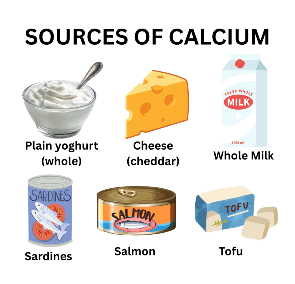
Best Food Sources of Calcium
Dietary calcium is always the preferred source. It carries no toxicity risk, comes with co-nutrients that enhance absorption, and does not carry the cardiovascular or kidney stone concerns associated with high-dose supplements.
|
Food Source |
Serving Size |
Calcium (mg) |
|
Plain yoghurt (whole) |
1 cup (245 g) |
296 mg ✦ Excellent source |
|
Cheese (cheddar) |
40 g (1.5 oz) |
303 mg |
|
Milk (whole) |
1 cup (240 ml) |
276–293 mg |
|
Sardines (canned, with bones) |
85 g (3 oz) |
325 mg |
|
Salmon (canned, with bones) |
85 g (3 oz) |
179 mg |
|
Tofu (set with calcium sulphate) |
½ cup (~126 g) |
Up to 861 mg. Check label |
|
Fortified plant milk (soy, almond, oat) |
1 cup (240 ml) |
300–450 mg (check label) |
|
Kale (raw) |
1 cup (~67 g) |
90 mg |
|
Broccoli (cooked) |
½ cup (~78 g) |
31 mg |
|
Almonds |
28 g (1 oz) |
76 mg |
|
White beans (cooked) |
½ cup (~90 g) |
96 mg |
|
Fortified orange juice |
1 cup (240 ml) |
~300 mg (check label) |
Table 8. Top dietary sources of calcium. Source: NIH ODS / USDA FoodData Central. Adult RDA = 1,000–1,200 mg/day.
🥛 Key Dietary Principle: Three servings of dairy daily (1 cup milk + 1 cup yoghurt + 40 g cheese) provides approximately 850–900 mg of highly bioavailable calcium, nearly the entire adult daily requirement. For non-dairy eaters, tofu set with calcium sulphate, fortified plant milks, and sardines with bones are the most practical alternatives.
Foods That Inhibit Calcium Absorption
- Oxalates: found in spinach, beet greens, and rhubarb. These bind calcium tightly, making it largely unabsorbable. Spinach calcium is only 5% bioavailable. Do not rely on spinach as a calcium source despite its calcium content.
- Phytates in whole grains and legumes reduce calcium absorption, though less dramatically than oxalates.
- Very high sodium diet: It increases urinary calcium excretion, a modifiable cause of negative calcium balance.
- Caffeine: modest effect. 1 cup of coffee reduces calcium retention by approximately 3 mg, clinically insignificant at normal intake levels.
Osteoporosis – The Long-Term Consequence of Calcium Deficiency
Osteoporosis is the most prevalent bone disease in the world. It is characterised by reduced bone mineral density, deteriorated bone microarchitecture, and dramatically increased fracture risk. It is the long-term consequence of sustained inadequate calcium and Vitamin D intake, combined with age-related bone loss and hormonal changes.
Risk Factors for Osteoporosis
- Female sex: women lose bone rapidly in the 5–10 years after menopause due to oestrogen deficiency
- Age: bone mass peaks at approximately age 30 and declines progressively thereafter
- Low lifetime calcium and Vitamin D intake
- Family history of osteoporosis or fragility fracture
- Smoking and excessive alcohol
- Corticosteroid use: the leading drug cause. It impairs calcium absorption and reduces bone formation
- Low body weight (BMI < 18.5)
- Hypogonadism, low oestrogen in women, low testosterone in men
- Chronic malabsorption conditions
Osteoporosis Treatment – Beyond Calcium
Adequate calcium and Vitamin D are necessary but not sufficient for treating established osteoporosis. Additional treatments for those at high fracture risk:
- Bisphosphonates (alendronate, risedronate): first-line, reduce fracture risk by 30–50%. Must be taken with water on an empty stomach, remaining upright for 30 minutes.
- Denosumab: monoclonal antibody against RANKL twice-yearly injection. Causes significant hypocalcaemia if Vitamin D is deficient. We must correct Vitamin D before starting.
- Teriparatide / abaloparatide: anabolic agents, stimulate new bone formation. Reserved for severe osteoporosis.
- Hormone replacement therapy (HRT): prevents post-menopausal bone loss, benefits must be balanced against individual risks.
How to Prevent Calcium Deficiency
For the General Population
- Aim for 3 servings of dairy daily or calcium-equivalent plant-based alternatives
- Include canned fish with bones (sardines, salmon) regularly
- Choose calcium-set tofu and fortified plant milks if dairy-free
- Maintain adequate Vitamin D levels. Without it, dietary calcium cannot be effectively absorbed
- Avoid very high sodium, excessive caffeine, and smoking – all impair calcium balance.
For High-Risk Groups
- Post-menopausal women: 1,200 mg calcium/day + 800–1,000 IU Vitamin D; DEXA scan at menopause and every 2 years thereafter
- Post-thyroidectomy patients: calcium and active Vitamin D (calcitriol) from day 1 post-surgery; daily calcium monitoring for first week
- Patients on long-term corticosteroids: 1,200–1,500 mg calcium + 800 IU Vitamin D daily; DEXA scan at baseline and annually
- Patients on PPIs > 1 year: use calcium citrate (not carbonate) absorbed without gastric acid; annual calcium monitoring
- Vegan and dairy-free individuals: plan calcium intake deliberately; use calcium-set tofu and fortified plant milks; consider supplement
- CKD patients: nephrologist-guided calcium and calcitriol management. Standard Vitamin D cannot be activated in CKD.
Calcium vs Magnesium vs Vitamin D Deficiency, How to Tell Them Apart
|
Feature |
Calcium Deficiency |
Magnesium Deficiency |
Vitamin D Deficiency |
|
Serum electrolyte affected |
Low serum calcium |
Low serum magnesium |
Low 25(OH)D |
|
Hallmark clinical sign |
Chvostek’s sign, Trousseau’s sign, tetany |
Nocturnal leg cramps, anxiety, arrhythmia |
Bone pain, muscle weakness, fatigue |
|
Cardiac risk |
QT prolongation, cardiac arrest in severe cases |
Torsade de pointes |
Arrhythmia – less direct |
|
Bone impact |
Osteoporosis (long-term) |
Contributes via Vitamin D activation failure |
Osteomalacia (adults), rickets (children) |
|
Critical rule |
Correct Mg first if refractory to treatment |
Correct before Ca or K can be fixed |
Needs Mg to be activated |
|
Key diagnostic test |
Adjusted serum calcium + PTH |
Serum magnesium (limited sensitivity) |
Serum 25(OH)D |
Table 9. Comparative guide — Calcium vs Magnesium vs Vitamin D deficiency. Source: Clinical experience + NCBI StatPearls; NIH ODS.
Frequently Asked Questions About Calcium Deficiency
Related Articles on MedBeaconHub.com
- Nutritional Deficiency Diseases: A Doctor’s Complete Guide (Pillar Article)
- Vitamin D Deficiency: The Most Common Cause of Dietary Calcium Deficiency
- Magnesium Deficiency: Why Correcting Magnesium Comes Before Calcium
- Vitamin K Deficiency: The Partner Mineral in Bone Health
- Potassium Deficiency: Another Electrolyte That Falls With Magnesium
- Iron Deficiency Anaemia: Complete Clinical Guide
References and Authoritative Sources
- NIH Office of Dietary Supplements — Calcium Fact Sheet for Health Professionals
- NCBI StatPearls — Hypocalcaemia
- Cleveland Clinic — Hypocalcaemia: Causes, Symptoms and Treatment
- Merck Manual Professional Edition — Hypocalcaemia
- Medscape — Hypocalcaemia: Background, Pathophysiology, Etiology
- UPMC — Hypocalcaemia: Causes, Symptoms and Treatments (Reviewed April 2025)
- Patient.info — Hypocalcaemia (Causes, Symptoms and Treatment)
- JAMA 2024 — Severe Hypocalcaemia with Denosumab Among Older Female Dialysis-Dependent Patients
- WHO — Osteoporosis and Musculoskeletal Conditions
- NIH ODS — Vitamin D Fact Sheet (for calcium absorption context)