
Copper Deficiency (Hypocupraemia): Symptoms, Causes, Myelopathy and Treatment
The trace mineral that powers your energy production, builds your collagen, makes your pigment, and whose deficiency can destroy your spinal cord in a pattern that looks exactly like Vitamin B12 deficiency — except the B12 is normal
✍️ Written and Reviewed by: Prof. Dr. Qazi Taqweemulhaq, FCPS Medicine. Professor of Medicine, Women Medical and Dental College, Abbottabad, Pakistan. Consultant Physician with 32 Years of Clinical Experience.📅 Last Updated: June 2026 | References: NIH ODS, NCBI StatPearls (2025), Merck Manual, Cleveland Clinic, Neurology, NEJM, American Journal of Clinical Nutrition
⚡ Quick Answer: Copper deficiency (hypocupraemia) is more common in clinical practice than most physicians realise. In the United States and Europe, the three most common causes are high-dose zinc supplementation (zinc induces intestinal metallothionein which traps copper and prevents its absorption), bariatric surgery, and malabsorption conditions including coeliac disease and Crohn’s disease. The hallmark clinical triad is a myelopathy indistinguishable from Vitamin B12 subacute combined degeneration, a normocytic or microcytic anaemia that does not respond to iron, and neutropenia. The adult RDA for copper is 900 mcg (0.9 mg) per day. Treatment is oral copper supplementation 2 mg daily, with the cause corrected simultaneously.
✅ KEY TAKEAWAYS — Copper Deficiency |
• Copper is required for at least eight critical enzymes including cytochrome c oxidase (energy production), superoxide dismutase (antioxidant defence), ceruloplasmin (iron metabolism), lysyl oxidase (collagen synthesis), and dopamine beta-hydroxylase (neurotransmitter production) |
• The most important and most commonly missed clinical presentation of copper deficiency is a progressive myelopathy that is clinically and radiologically identical to Vitamin B12 subacute combined degeneration — with normal B12 levels. Any patient with suspected B12 myelopathy and a normal or borderline B12 must have serum copper and ceruloplasmin checked |
• High-dose zinc supplementation is the most common cause of copper deficiency in contemporary American clinical practice. Zinc above 50 mg per day induces intestinal metallothionein, which binds copper preferentially and prevents its absorption. Millions of Americans take zinc supplements for immune support at doses that silently deplete copper over months |
• Copper deficiency causes an anaemia that does not respond to iron supplementation because ceruloplasmin (a copper-dependent protein) is required to oxidise ferrous iron for transport and incorporation into haemoglobin |
• Menkes disease is a fatal X-linked genetic copper deficiency disorder presenting in male infants with kinky hair, neurological degeneration, and connective tissue failure. Early IV copper treatment is partially effective if started before irreversible neurological damage |
• The diagnostic tests are serum copper (below 70 µg/dL indicates deficiency) and serum ceruloplasmin (below 20 mg/dL) alongside a 24-hour urine copper to assess body copper balance |
The Man Whose B12 Was Normal
He was 58 years old. Progressive difficulty walking for two years. His gait had become uncertain and wide-based. He was dropping objects. His hands were clumsy. He had fallen twice in the past three months.
A neurologist reviewed him and documented loss of vibration sense in both feet, absent ankle reflexes, a positive Romberg sign, and brisk knee reflexes with upgoing plantar responses. The picture was classic posterior and lateral column involvement. Subacute combined degeneration of the spinal cord.
The team checked Vitamin B12. It was 310 pg/mL. Normal.
They checked it again with a methylmalonic acid (MMA) level. Also normal.
The patient was admitted to neurology. MRI of the cervical and thoracic spine showed the characteristic posterior column signal changes seen in B12 myelopathy. But the B12 was unequivocally normal.
A junior doctor remembered reading about copper deficiency myelopathy during a recent journal review. Serum copper was checked. It was 42 µg/dL — profoundly low. Ceruloplasmin was undetectable.
When the history was reviewed more carefully, one detail had been missed in the initial clerking. For two years — exactly coinciding with the start of his neurological symptoms — he had been taking zinc acetate 150 mg daily. He had read online that high-dose zinc prevents viral infections. He had never been told that zinc at this dose depletes copper.
He was started on oral copper supplementation 2 mg daily and his zinc was stopped. Over eight months his gait partially improved. His vibration sense returned to his knees but not his feet. Some of the damage was irreversible.
🏥 From My Clinic: I now check serum copper in every patient with a myelopathy or peripheral neuropathy where B12 is normal or borderline. I also check it in every patient on long-term high-dose zinc supplements. Copper deficiency myelopathy is one of the most under-diagnosed neurological conditions in Western clinical practice. It is not rare. It is simply not thought of. For the complete guide to nutritional deficiency diseases, read: Nutritional Deficiency Diseases: The Complete Doctor’s Guide.
What Copper Does
Copper is an essential trace mineral that serves as a cofactor for at least eight critical enzymes in human physiology. The body contains approximately 100 to 150 mg of total copper, distributed primarily in skeletal muscle (approximately 38%), brain (approximately 11%), liver (approximately 15%), and bone. The liver is the primary organ of copper storage and metabolism.
The Eight Copper-Dependent Enzymes
Cytochrome c oxidase (Complex IV). The terminal enzyme of the mitochondrial electron transport chain. Copper is essential for the transfer of electrons to oxygen to generate ATP. Without adequate copper, aerobic energy production in every cell is impaired. This is why copper deficiency produces profound fatigue alongside its neurological and haematological consequences.
Superoxide dismutase (Cu/Zn-SOD). The primary antioxidant enzyme that neutralises the superoxide radical, converting it to hydrogen peroxide which is then disposed of by catalase. Copper deficiency impairs antioxidant defence in a way that parallels selenium deficiency (which impairs glutathione peroxidase). Both minerals protect cells from oxidative damage through different enzymatic routes.
Ceruloplasmin. The primary copper transport protein in plasma. But ceruloplasmin also functions as a ferroxidase — it oxidises ferrous iron (Fe²⁺) to ferric iron (Fe³⁺), which is required for iron to bind transferrin and be transported to developing red blood cells. Without ceruloplasmin, iron accumulates in tissues (particularly the liver and brain) but cannot be mobilised for haemoglobin synthesis. This is why copper deficiency produces an anaemia that does not respond to iron supplementation — the iron is present but trapped.
Lysyl oxidase. Catalyses the cross-linking of collagen and elastin fibres. This cross-linking gives connective tissue, blood vessel walls, and bone matrix their tensile strength. In copper deficiency, collagen and elastin are structurally weak. Blood vessels become fragile. Bones become brittle. In Menkes disease — the genetic form of copper deficiency — the characteristic tortuous, fragile arteries that develop reflect lysyl oxidase failure.
Dopamine beta-hydroxylase. Converts dopamine to norepinephrine. Copper deficiency impairs this conversion, reducing norepinephrine production. This is one mechanism by which copper deficiency produces neuropsychiatric symptoms including depression, anxiety, and autonomic dysfunction.
Tyrosinase. The enzyme required for melanin synthesis. Copper deficiency impairs melanin production, causing depigmentation of skin and hair. This is strikingly visible in Menkes disease, where the characteristic pale, kinky hair directly reflects tyrosinase failure from copper deficiency.
Peptidylglycine alpha-amidating monooxygenase (PAM). Required for the activation of many neuropeptides including vasopressin, oxytocin, and various GI hormones. PAM deficiency from copper insufficiency contributes to neurological and endocrine dysfunction.
Clotting factor activation. Copper participates in the synthesis of clotting factors. Copper deficiency can produce a mild bleeding tendency alongside the more prominent anaemia and neutropenia.
How Much Copper Do You Need Per Day
Group | RDA (mcg per day) | Upper Limit (mcg per day) |
Adults 19 years and over | 900 mcg (0.9 mg) | 10,000 mcg (10 mg) |
Pregnant women | 1,000 mcg | 10,000 mcg |
Breastfeeding women | 1,300 mcg | 10,000 mcg |
Children 1 to 3 years | 340 mcg | 1,000 mcg |
Children 4 to 8 years | 440 mcg | 3,000 mcg |
Children 9 to 13 years | 700 mcg | 5,000 mcg |
Table 1. Copper RDA and Tolerable Upper Intake Levels. Source: NIH ODS. The upper limit of 10 mg per day refers to all sources. Chronic intake above 10 mg per day causes hepatotoxicity and liver failure.
Copper Status in the United States and Europe
Dietary copper deficiency from food alone is uncommon in adults eating a varied US or EU diet. Oysters, beef liver, dark chocolate, nuts, and seeds provide ample copper in a mixed diet. NHANES data shows mean copper intakes in US adults of approximately 1.1 to 1.6 mg per day — above the RDA.
The clinically significant copper deficiency encountered in the US and EU almost always has a specific cause beyond inadequate dietary intake: zinc supplementation, malabsorption, bariatric surgery, or premature birth. This makes copper deficiency a condition of clinical contexts, not simply of dietary poverty.
Symptoms of Copper Deficiency
The symptoms of copper deficiency reflect its roles in haematopoiesis, neurological function, immune defence, and connective tissue maintenance. The presentation varies considerably depending on whether the deficiency is acute and severe (as in premature neonates receiving copper-free parenteral nutrition) or chronic and gradually progressive (as in adults with long-term zinc supplementation or post-bariatric malabsorption).
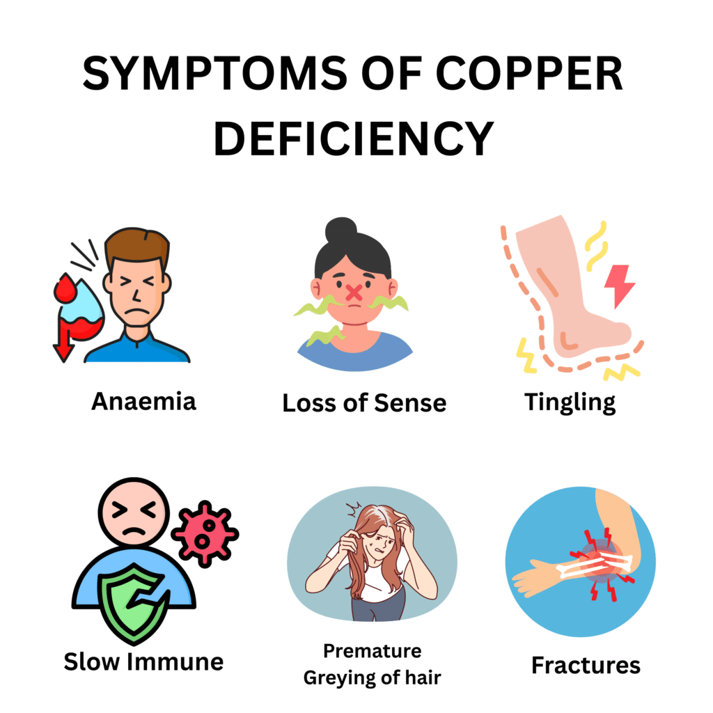
System | Mild–Moderate Deficiency | Severe / Prolonged Deficiency |
Blood | Normocytic or microcytic anaemia that does not respond to iron. Neutropenia (low white blood cell count). Occasionally thrombocytopenia | The anaemia is the most common presenting laboratory finding. It is indistinguishable from iron deficiency on the blood count but serum ferritin and iron are normal or elevated |
Neurological — spinal cord | Progressive myelopathy: loss of vibration sense and proprioception, Romberg sign positive, brisk reflexes, upgoing plantars. MRI shows posterior and lateral column T2 signal changes identical to B12 subacute combined degeneration | This is the most serious neurological feature. Onset is insidious. May progress over months to years before diagnosis |
Neurological — peripheral nerves | Peripheral neuropathy: tingling, numbness, and burning in the hands and feet. Length-dependent sensorimotor pattern | Coexists with myelopathy in many cases. Contributes to the clinical picture alongside the posterior column involvement |
Neurological — optic nerve | Optic neuropathy in severe cases: progressive visual loss, optic atrophy on fundoscopy | Less common but documented. Responds partially to copper replacement if caught early |
Immune | Neutropenia: recurrent bacterial infections, slow recovery from illness | The neutropenia of copper deficiency closely resembles that of drug-induced or autoimmune neutropenia. Bone marrow shows vacuolation of myeloid precursors |
Connective tissue | Osteoporosis and fractures in chronic deficiency. Skin laxity. Vascular fragility | Lysyl oxidase failure weakens collagen cross-links |
Skin and hair | Premature greying of hair. Depigmentation in severe deficiency. Pale skin | Tyrosinase failure reduces melanin production |
Cardiovascular | Anaemia-related cardiovascular strain. In severe genetic deficiency: aortic aneurysm and vascular rupture | Lysyl oxidase failure weakens aortic wall in Menkes disease |
Table 2. Symptoms of copper deficiency. Sources: NIH ODS; NCBI StatPearls (2025); Merck Manual; Neurology (2024 Review).
The Copper Deficiency Myelopathy — The Diagnosis Physicians Miss
This is the most clinically significant and most commonly missed feature of copper deficiency in adult patients. It deserves its own detailed discussion because the consequences of missing it are severe and often irreversible.
Copper deficiency myelopathy affects the dorsal (posterior) columns and lateral corticospinal tracts of the spinal cord — exactly the same anatomical distribution as Vitamin B12 subacute combined degeneration. The clinical signs are therefore identical: loss of vibration sense beginning in the feet, impaired proprioception, positive Romberg sign, brisk deep tendon reflexes, and upgoing plantar responses (Babinski sign). MRI of the spine shows characteristic T2 hyperintensity in the posterior columns.
The single critical difference: the B12 level is normal.
When a patient presents with this clinical and radiological picture and B12 is normal or borderline, most neurologists will check MMA and homocysteine to confirm true B12 deficiency. If these are also normal, the diagnosis is unclear. Many patients at this point are labelled as having idiopathic myelopathy, degenerative spine disease, or multiple sclerosis — and the actual treatable cause goes unidentified for months or years.
The correct next step when B12 myelopathy is suspected but B12 is normal: check serum copper and ceruloplasmin. This single test takes minutes to order and dramatically changes the clinical trajectory for the patient whose copper is low.
💡 Clinical Insight: I have learned to think of copper deficiency and B12 deficiency as the two treatable causes of posterior column myelopathy in adults. When one is normal, always check the other. Do not stop at a normal B12. Do not stop at a normal copper. Check both simultaneously.
The Anaemia That Looks Like Iron Deficiency But Isn’t
The anaemia of copper deficiency is characteristically normocytic or microcytic with normal or elevated serum ferritin and transferrin saturation — the opposite of true iron deficiency where ferritin is low and transferrin saturation is reduced.
The mechanism: ceruloplasmin (a copper-dependent ferroxidase) is required to oxidise Fe²⁺ to Fe³⁺ so that iron can bind transferrin for transport to erythroid precursors in the bone marrow. Without ceruloplasmin activity, iron accumulates in tissues but cannot be mobilised for haemoglobin synthesis.
The practical consequence: a patient with copper deficiency anaemia who is given iron supplementation shows no improvement, because iron is not the limiting factor. It is already present. The limiting factor is the failure of copper-dependent iron transport.
When an anaemia does not respond to iron after 6 to 8 weeks of adequate supplementation, copper deficiency is on the differential. Check serum copper and ceruloplasmin alongside B12, folate, and riboflavin — all of which can cause anaemia resistant to iron therapy.
What Causes Copper Deficiency
Cause | Mechanism | Who Is Most Affected |
High-dose zinc supplementation | The most important and most common cause in contemporary US and EU practice. Zinc at doses above 50 mg per day induces metallothionein, a metal-binding protein, in intestinal enterocytes. Metallothionein has a higher binding affinity for copper than for zinc. Dietary copper binds metallothionein, becomes trapped inside intestinal cells, and is excreted when those cells shed — never reaching the circulation. This occurs silently over months to years | Americans taking zinc supplements for immune support at 50 to 200 mg per day. This practice became dramatically more widespread following pandemic-era supplementation advice. Many US supplement products contain 50 mg zinc per capsule — more than sufficient to deplete copper with daily use |
Bariatric surgery | Gastric bypass surgery reduces gastric acid production and bypasses the duodenum and proximal jejunum — the primary sites of copper absorption. The acidic environment of the stomach normally releases copper from food and prepares it for absorption. After bypass, this step is impaired | Post-Roux-en-Y gastric bypass patients are at the highest risk. Copper is one of the trace minerals most consistently depleted after bariatric surgery, alongside iron, zinc, and B12. Lifelong monitoring and supplementation are required |
Coeliac disease | Villous atrophy from untreated coeliac disease reduces absorptive surface area throughout the small intestine, impairing absorption of multiple trace minerals including copper | Any patient with coeliac disease, particularly those with incomplete mucosal recovery on a gluten-free diet. Should be monitored annually alongside iron, folate, zinc, and Vitamin D |
Crohn’s disease | Active intestinal inflammation impairs copper absorption. Significant small bowel involvement is most relevant | Patients with extensive Crohn’s disease, particularly those with jejunal involvement |
Total parenteral nutrition without copper | Historically, TPN formulations omitted copper. Modern TPN includes copper as standard but home TPN and non-standard formulations remain a risk | Patients on home TPN or non-standard supplementation. A fully preventable cause |
Excessive antacid use | Antacids neutralise gastric acid. Copper absorption is acid-dependent. Chronic antacid use impairs copper bioavailability from food | Patients on high-dose antacid regimens, particularly those also on proton pump inhibitors long-term |
Premature birth | Copper crosses the placenta primarily in the third trimester. Premature infants miss this critical transfer and are born with minimal copper stores. Breast milk is low in copper. Without supplementation, severe copper deficiency develops rapidly | Premature infants especially those below 32 weeks gestation. Standard neonatal nutritional protocols must include copper |
Chronic kidney disease | CKD stages 3–5, dialysis patients | Second hydroxylation (25(OH)D → calcitriol) fails in CKD. Active calcitriol is needed |
Menkes disease (genetic) | X-linked recessive mutation in ATP7A — the copper transport ATPase that moves copper from intestinal cells into the circulation. Copper is absorbed into enterocytes but cannot be exported. It accumulates in intestinal cells while the rest of the body is profoundly deficient | Male infants exclusively (X-linked). Presents in the first weeks of life. Incidence approximately 1 in 100,000 to 250,000 live male births |
Malnutrition and extreme dietary restriction | Very low intake of animal proteins, nuts, and seeds combined with very low total caloric intake | Patients with severe anorexia nervosa, those on extremely restricted diets, and individuals with food insecurity |
Table 3. Causes of copper deficiency. Sources: NIH ODS; NCBI StatPearls (2025); Merck Manual; Neurology.
The Zinc-Copper Interaction — The Most Important Public Health Message
This interaction is the most clinically important aspect of copper deficiency in the contemporary US and EU context, and it is almost entirely unrecognised by the public.
The mechanism is well established in biochemistry. Zinc induces the synthesis of metallothionein in intestinal enterocytes. Metallothionein is a cysteine-rich protein with an extraordinarily high affinity for heavy metals — including both zinc and copper. When metallothionein is induced by zinc, dietary copper binds it tightly within intestinal cells. When those cells complete their 3 to 5 day lifespan and shed into the gut lumen, the copper bound within them is lost in the stool. None of it is absorbed.
The clinical consequence: a person taking zinc 150 mg daily is inducing enough metallothionein to trap essentially all dietary copper from the intestine. The process is silent. Symptoms of copper deficiency — anaemia, neutropenia, and eventually myelopathy — develop over months to years while the patient continues taking their zinc supplement in the belief that they are supporting their immune health.
In the United States, zinc supplements of 50 to 200 mg per tablet are widely sold without prescription and actively marketed for immune support. The recommended dietary allowance for zinc is 8 to 11 mg per day for adults. These supplements provide 5 to 18 times the RDA in a single daily tablet. Many patients combine multiple supplements that each contain zinc, inadvertently consuming 100 mg or more per day for extended periods.
The threshold for copper depletion: doses of zinc above approximately 50 mg per day taken consistently for months will deplete copper stores in most adults. The higher the dose and the longer the duration, the more severe the depletion.
The clinical rule: any adult taking zinc supplements above 25 to 30 mg per day chronically requires either copper co-supplementation (1 to 2 mg copper per 50 mg zinc) or regular monitoring of serum copper and ceruloplasmin. This rule is almost never communicated on zinc supplement labels or by the practitioners who recommend them.
Wilson disease (copper overload) is the opposite problem — ATP7B gene mutation causing copper accumulation rather than deficiency. It is not discussed in this article but is mentioned here to clarify: copper supplementation is contraindicated in Wilson disease patients.
Menkes Disease — The Fatal Genetic Form
Menkes disease is the most severe form of copper deficiency and represents the complete opposite end of the spectrum from dietary or drug-induced adult deficiency.
The gene affected is ATP7A, which encodes the copper-transporting ATPase responsible for moving copper from intestinal enterocytes into the portal circulation and for distributing copper within cells to copper-dependent enzymes. When ATP7A is mutated, copper cannot be exported from intestinal cells. It accumulates in the gut while the brain, liver, and every other organ is profoundly deprived.
The clinical presentation is distinctive and heartbreaking:
Kinky or steely hair (pili torti): The hair is sparse, brittle, and twisted under microscopy. This reflects failure of the disulfide bonds in keratin that require copper-dependent enzyme activity. The hair abnormality is a direct and pathognomonic physical sign.
Progressive neurological degeneration: Seizures, profound hypotonia, and rapid developmental regression beginning in the first weeks of life. The brain requires copper at every level of development and degenerates rapidly without it.
Connective tissue failure: Bladder diverticula, joint laxity, skin that is pale, loose, and doughy. The tortuous, fragile arteries produced by lysyl oxidase failure can rupture, causing subdural haematoma and stroke-like events.
Hypothermia: Copper-dependent thermogenesis fails.
Without treatment, Menkes disease is fatal, usually before the age of three. Early intravenous or subcutaneous copper histidine therapy, initiated before irreversible neurological damage has occurred (ideally within the first few weeks of life), can partially stabilise or improve the neurological outcome. Newborn screening programmes in some US states now include Menkes disease to enable this early intervention, though it is not universally screened.
Diagnosis
Copper deficiency is a laboratory diagnosis in most adult patients, confirmed by the combination of low serum copper and low ceruloplasmin in the appropriate clinical context.
Blood Tests
Serum copper: The primary diagnostic test. Reference range: 70 to 140 µg/dL (11 to 22 µmol/L) in most US and European laboratories. Below 70 µg/dL indicates deficiency. Below 50 µg/dL indicates severe deficiency.
Important caveat: serum copper is an acute-phase reactant. It rises during infection, inflammation, pregnancy, and oestrogen use (including the oral contraceptive pill). A patient with an inflammatory illness may have a falsely normal or even elevated serum copper despite true tissue copper depletion. Interpret results in the context of inflammatory markers (CRP, ESR).
Serum ceruloplasmin: The most clinically useful marker for copper status. Ceruloplasmin is synthesised in the liver and requires copper for its structure and function. It falls reliably in copper deficiency. Reference range: 20 to 35 mg/dL. Below 20 mg/dL indicates copper deficiency. Undetectable ceruloplasmin indicates severe deficiency or aceruloplasminaemia (a rare genetic condition).
Like serum copper, ceruloplasmin is also an acute-phase reactant — it rises in inflammation. Both must be interpreted together. Low ceruloplasmin in the context of elevated CRP is highly significant for true copper deficiency.
24-hour urine copper: Reflects copper excretion and helps assess body copper status and balance. In copper deficiency, urinary copper is typically very low (below 15 µg per 24 hours). Used alongside serum levels for complete assessment. Also useful in Wilson disease monitoring where it is elevated.
Complete blood count: Anaemia (normocytic or microcytic), neutropenia, and occasionally thrombocytopenia. The combination of anaemia plus neutropenia with normal or elevated serum ferritin strongly suggests copper deficiency.
Serum ferritin and iron studies: To confirm that the anaemia is not primary iron deficiency. In copper deficiency, ferritin is normal or elevated and transferrin saturation is normal, distinguishing it from iron deficiency anaemia.
Vitamin B12 and methylmalonic acid: Always check simultaneously with copper in any myelopathy workup. B12 myelopathy and copper myelopathy can coexist. Both must be addressed.
Serum zinc: Zinc excess drives copper depletion. In patients taking high-dose zinc, serum zinc may be elevated or normal but the history is the key diagnostic clue.
MRI spine: In patients with myelopathy, MRI of the cervical and thoracic spine shows characteristic T2 hyperintensity in the posterior columns — identical to B12 myelopathy and not distinguishable on imaging alone. The clinical laboratory values are what differentiate the two.
Bone marrow examination: If the diagnosis remains uncertain, bone marrow aspirate in copper deficiency shows characteristic vacuolation of myeloid and erythroid precursors — a morphological pattern that strongly suggests the diagnosis when present alongside low serum copper.
Treatment
Clinical Scenario | Treatment | Duration and Notes |
Zinc-induced copper deficiency | Stop high-dose zinc immediately. Oral copper gluconate or copper sulphate 2 mg daily | Continue copper replacement until serum copper and ceruloplasmin normalise (typically 4 to 8 weeks). If long-term zinc is needed for a medical reason, balance with 1 to 2 mg copper per 50 mg zinc |
Post-bariatric surgery copper deficiency | Oral copper 2 mg daily as part of comprehensive post-bariatric micronutrient protocol. Water-miscible forms preferred | Lifelong supplementation and annual serum copper monitoring mandatory. Include alongside iron, zinc, B12, and Vitamin D supplementation |
Malabsorption-related deficiency (coeliac, Crohn) | Oral copper 2 to 4 mg daily. Address underlying condition simultaneously | Treat the malabsorption cause. Copper requirement may remain elevated if absorption is chronically impaired |
Copper deficiency myelopathy | IV copper 2 to 4 mg daily for 5 to 7 days initially in severe neurological involvement, then oral copper 2 mg daily maintenance | Neurological improvement is partial and slow — months to a year. Earlier treatment = better outcome. IV route used when absorption is uncertain or disease is severe |
Anaemia and neutropenia | Oral copper 2 mg daily. Do not give iron unless iron deficiency is separately confirmed | Blood count normalises within 4 to 8 weeks of copper replacement |
Total parenteral nutrition patients | Copper 0.3 to 0.5 mg per day added to TPN formulation as standard | Mandatory inclusion in all TPN. Adjust for biliary losses (increase in cholestasis) and inflammatory state |
Menkes disease | IV or subcutaneous copper histidine as early as possible after diagnosis | Lifelong. Early initiation (before irreversible neurological damage) improves outcome. Specialist metabolic team management essential |
Premature neonates | Copper supplementation as part of standard preterm infant nutritional protocol | Follow neonatal guidelines. Breast milk is supplemented. TPN must include copper |
Table 4. Copper deficiency treatment protocol. Sources: NIH ODS; NCBI StatPearls (2025); Merck Manual; Neurology.
Recovery Timeline
What Improves | Timeline |
Serum copper normalises | 4 to 8 weeks with oral supplementation |
Neutropenia corrects | 4 to 8 weeks |
Anaemia corrects | 6 to 12 weeks |
Peripheral neuropathy improves | 3 to 6 months |
Myelopathy — early cases | Partial improvement over 6 to 12 months |
Myelopathy — established severe cases | Minimal or no improvement. Prevention and early diagnosis are essential |
Table 5. Copper deficiency recovery timeline. Source: Clinical experience; NCBI StatPearls (2025); Neurology.
The critical message about the myelopathy: Neurological recovery from copper deficiency myelopathy is partial at best and often disappointing. The posterior column damage is structural and slow to regenerate. In most reported series, patients improve somewhat — gait becomes safer, vibration sense returns partially — but complete recovery is unusual. Early diagnosis, before significant structural cord damage has accumulated, is the only way to preserve full neurological function. This is why the clinical index of suspicion must be high in any patient with risk factors.
Best Food Sources of Copper
The copper content of food is highly variable. Animal organ meats — particularly beef liver and oysters — are by far the richest sources. Most plant foods contain moderate amounts of copper, with the notable exceptions of nuts, seeds, and dark chocolate.

Food | Serving | Copper (mcg) | Notes |
Oysters, cooked | 3 oysters (85 g) | 4,850 mcg | The single richest food source by a very large margin. Five times the daily RDA per serving |
Beef liver, cooked | 85 g | 12,400 mcg | |
Shiitake mushrooms, cooked | Half cup | 650 mcg | Best plant copper source per serving |
Dark chocolate (70 to 85%) | 28 g (1 oz) | 500 mcg | Practical everyday source |
Cashews | 28 g | 622 mcg | Excellent nut source |
Sunflower seeds | 28 g | 519 mcg | Good plant source alongside Vitamin E and selenium |
Sesame seeds (tahini) | 2 tablespoons | 730 mcg | Outstanding plant source |
Almonds | 28 g | 333 mcg | Good everyday nut |
Black beans, cooked | Half cup | 180 mcg | |
Lentils, cooked | Half cup | 250 mcg | |
Tofu, raw | Half cup | 476 mcg | Good plant source |
Potato, baked with skin | 1 medium | 167 mcg | Modest but practical daily source |
Salmon, cooked | 85 g | 273 mcg |
Table 6. Top dietary sources of copper. Source: NIH ODS; USDA FoodData Central. Adult RDA: 900 mcg (0.9 mg) per day.
The zinc-copper dietary balance: The dietary ratio of zinc to copper matters as much as absolute intake of either mineral. The optimal dietary zinc-to-copper ratio is approximately 8 to 12:1. Most Western diets naturally provide this ratio when eaten as whole foods. The problem arises exclusively with supplementation at non-physiological doses. A person eating oysters, liver, dark chocolate, nuts, and seeds alongside their normal diet is not at risk of copper deficiency from food alone. The risk is entirely in the supplement context.
Frequently Asked Questions About Copper Deficiency
Related Articles on MedBeaconHub.com
- Nutritional Deficiency Diseases: The Complete Doctor’s Guide
- Zinc Deficiency: Immunity, Taste and the Copper Connection
- Vitamin B12 Deficiency: Nerve Damage and Pernicious Anaemia
- Iron Deficiency Anaemia: Symptoms and Treatmen
- Selenium Deficiency: The Thyroid and Mitochondrial Mineral
- Magnesium Deficiency: Sleep, Anxiety and Muscle Cramps
- Folate Deficiency: The Pregnancy-Critical B Vitamin
- Vitamin D Deficiency: The Most Common US Deficiency
- Calcium Deficiency: Hypocalcaemia and Bone Disease
- Vitamin B1 Thiamine Deficiency: Beriberi and Wernicke Encephalopathy
- Vitamin B2 Riboflavin Deficiency: Symptoms, Migraine and MTHFR
- Vitamin B3 Niacin Deficiency: Pellagra and the Four Ds
- Vitamin B6 Deficiency: Depression, Neuropathy and the OCP
- Vitamin A Deficiency: Night Blindness and Immunity
- Vitamin K Deficiency: Bleeding and Warfarin
References and Authoritative Sources
- NIH Office of Dietary Supplements — Copper Fact Sheet for Health Professionals
- NCBI StatPearls — Copper Deficiency (Updated 2025)
- Merck Manual Professional Edition — Copper Deficiency
- Cleveland Clinic — Copper Deficiency
- Neurology — Copper Deficiency Myelopathy: A Systematic Review (2024)
- American Journal of Clinical Nutrition — High-Dose Zinc Supplementation and Copper Status (2024)
- PMC — Copper Deficiency After Bariatric Surgery: Systematic Review (2024)
- Genetics in Medicine — Menkes Disease: Diagnosis, Management, and Outcomes (2023)
- PMC — Copper Deficiency Myelopathy and Zinc Supplementation (2024)
- NEJM — Copper Deficiency Myeloneuropathy (2023)